Digital Forensics
The abundance of forensic analysis in today’s television programmes rarely touches the role of modern technology and engineering. While there is still a role for the microscope in forensic work, research teams at Warwick Manufacturing Group (WMG) at the University of Warwick have shown that laser scanning, computer tomography, 3D printing and advanced 3D visualisation can collect and analyse evidence for crime investigation teams.
The non-invasive nature of these technologies is ideal for recording and documenting evidence, for analysis and making presentations to juries. Working with pathologists and staff at University Hospital Coventry and Warwickshire NHS Trust, WMG has helped crime scene investigators and the West Midlands Police in a number of homicide investigations and successful prosecutions.
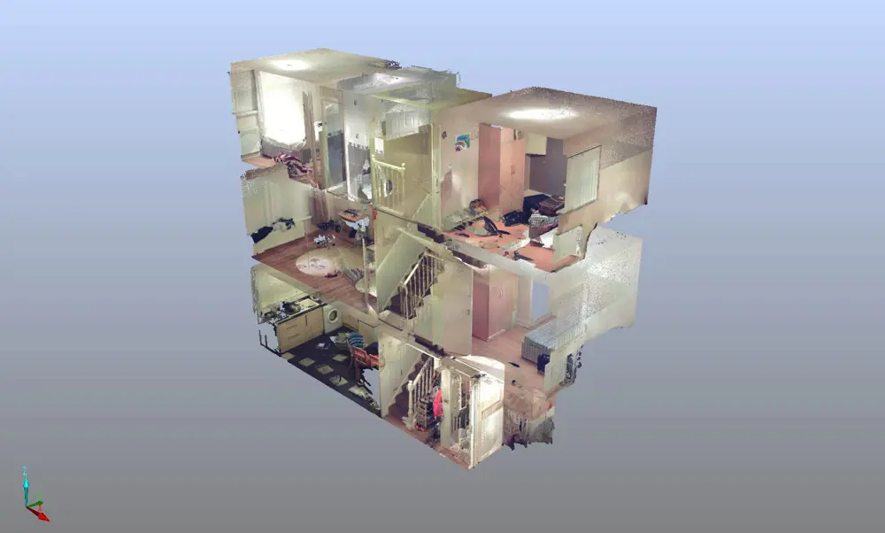
3D representation of a complex multiple-room crime scene © West Midlands Police
Crime scene
The forensic process starts at crime scenes that can present complex scenarios that require analysing many rooms over several storeys. There can be hundreds of potential items of interest at the scene. Evidence such as weapons, blood splatter, fibres, fingerprints, DNA and footprints need to be collected, documented and analysed.
The forensic analysis on TV usually starts with a photographer recording the scene and taking standard high-resolution 2D photos with a digital SLR camera. The WMG team has now taken this to a new level. A typical crime scene can generate hundreds of detailed images. The challenge comes when a case goes to trial and the prosecution has to communicate the narrative of the crime and sequencing of events to a jury who might not visit the location.
Cameras and tape measures and other equipment can record additional data, but this can be time consuming and sometimes unreliable. There is also the challenge that crimes can take place in public amenities or commercial areas where there could be pressure to release a property back to its owner or to reopen a facility. It can cost many thousands of pounds a day to lock down scenes in these situations.
3D scene-scanning technology is used for the investigation of crimes, accidents and fires. This technique was originally developed for architectural and geological surveying and is commonly used for construction and civil engineering applications.
3D laser scanning captures millions of data points, using a Cartesian coordinate system that help to document and model a real-world environment. The result is a 3D ‘point cloud’ that can be used to recreate a virtual model for future analysis.
An operator can collect over 10 million data points in less than five minutes, capturing the geometric characteristics of a specific location to form a 360° laser scan of an indoor scene with accuracies within 2 mm, up to a distance of 330 m
An operator can collect over 10 million data points in less than five minutes, capturing the geometric characteristics of a specific location to form a 360° laser scan of an indoor scene with accuracies within 2 mm, up to a distance of 330 m. This technology relies on line-of-sight imaging, so it usually requires multiple scans from different angles, taking the total cloud count number to over 100 million individual points. This point cloud can then be exported into a software package that combines the individual scans into a single data set. The software can then process the data and render it into a 3D model of the location. In this way 3D laser scanning can create a permanent virtual archive of the scene.
This ‘virtual crime scene’ is just the beginning of the forensic analysis. For example, it can also be combined with physical measurements taken to support blood-pattern analysis, to corroborate line-of-sight for witness statements, quantification of critical distances and calculations of trajectories. Scanned data also make it possible to retrospectively revisit the scene of the crime as it was, long after the investigators have left the location. Investigating teams can also embed digital hyperlinks into these digital models, linking to additional detailed evidence collected from the crime scene. This means that the investigators can collate 2D photos, electronic copies of reports and audio, as well as video files. Such capability makes it possible to present valuable contextual information to a jury at court.
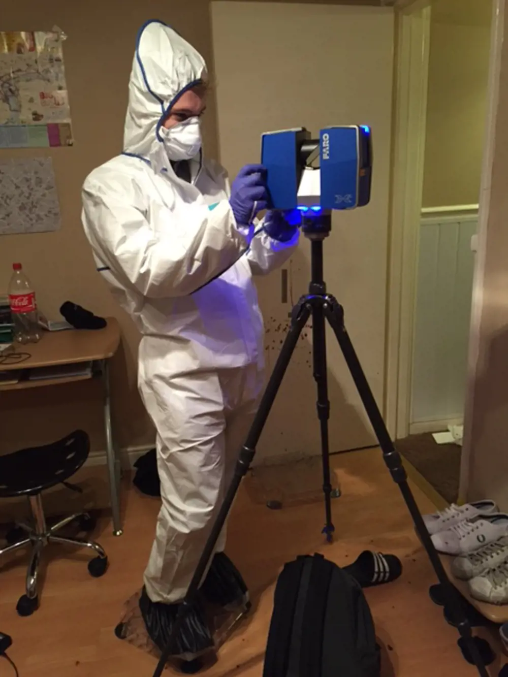
WMG Research Engineer Dan Norman setting up a 3D scene scanner in a live crime scene environment, wearing full forensic personal protective equipment
The technology of 3D imaging and the ability to generate a 3D virtual crime scene was used to great effect in 2015 with a murder investigation in a multi-storey domestic property in Birmingham. The crime took place throughout a number of different rooms. The ability to see cross sections and close ups of various rooms and corridors linked together in context, rather than a hundred different photos, helped the successful prosecution case.
As well as representing the physical characteristics of a crime scene, it helps to have virtual models that are as visually realistic as possible. However, point-cloud data contains limited information on texture and colour, yet this needs to be included within the model if it is to be realistic. The standard technique used to achieve this is to overlay high-resolution colour digital images onto the point cloud in the form of a 360° mosaic. This can be achieved manually with a digital SLR camera or using a scanner with an integrated charged-couple device chip that captures colour images as part of the scanning process. This image data can then encode important colour information onto each individual point in the cloud, helping to render a photorealistic result.
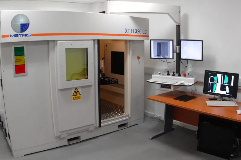
CT setup
🦴 How does a CT scan generate an image?
A typical computed tomography setup consists of a powerful X-ray source, a high-definition detector, with typically 2,000 by 2,000 pixels, and a manipulator for rotating the sample in the X-ray beam. As X-rays interact with the object, they are attenuated or pass through and arrive at the detector. The proportion of X-ray energy absorbed during the attenuation process results in a 2D radiograph image of the object where the grey value at each individual point depends on the amount and type of material that the X-rays have interacted with as they pass through the specimen. The object is rotated through 360° in the X-ray beam, taking over 3,000 individual radiographs at precise angular increments.
Mathematical CT technique can then reconstruct these radiographs to generate a single 3D model. Using X-rays makes it possible to capture all interior features in 3D at a microscopic level, highlighting details that are not visible to the human eye and that conventional medical scanning techniques would have missed.
WMG currently utilises two micro-CT scanners to support its forensic activities. In addition to the scanning technology, specialist software allows WMG to carry out the detailed analysis of the CT data and produce reports.
Working on sensitive criminal cases requires documentation and careful adherence to procedures that are not normal for academic research groups. For example, a Home Office pathologist or senior investigative officer can raise a formal request to carry out a micro-CT scan if, during a post mortem or autopsy, additional investigation is required.
WMG’s micro-CT scanners enable cone beam X-ray sources ranging from 160 kV–320 kV, a voxel special resolution down to sub-micron levels (170 nm, 1 x 10⁻⁹ m) , and a maximum sample size of 300 mm in diameter
3D scanning
Other techniques can add to the information, and to the realism, of virtual crime scenes. For example, WMG has supported West Midlands Police in integrating high dynamic range imaging (HDRi) technology into data collection. Standard digital photography is designed to capture light that is available to the human eye and captures only a limited range of light luminosity. This relatively narrow exposure range means that the images can obscure bright lighting or dark shadows, leading to their omission from the data set. Using camera systems with HDRi technology represents a step change in the ability to capture more accurate images of the light scene.
Although the benefits of 3D scene scanning to the criminal justice process are clear, there are challenges to its widespread implementation. Laser scanners are relatively cheap, but turning a large point cloud into a rendered model with fully immersive and interactive capabilities remains a lengthy and complex process. As well as the proprietary software used to process the point cloud, the analysis also requires additional software to transform the data into a fully immersive virtual environment that works at a practical level. This requires specialist skills and powerful computing hardware that would normally be financially prohibitive to law enforcement agencies. Such expertise, therefore, needs to be subcontracted or made available through a strategic partnership, as has been established between the University of Warwick and West Midlands Police.
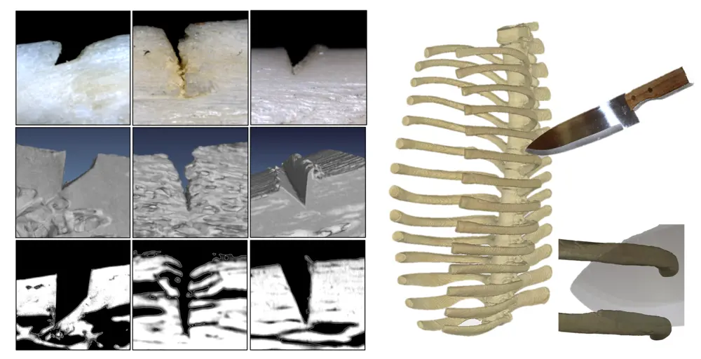
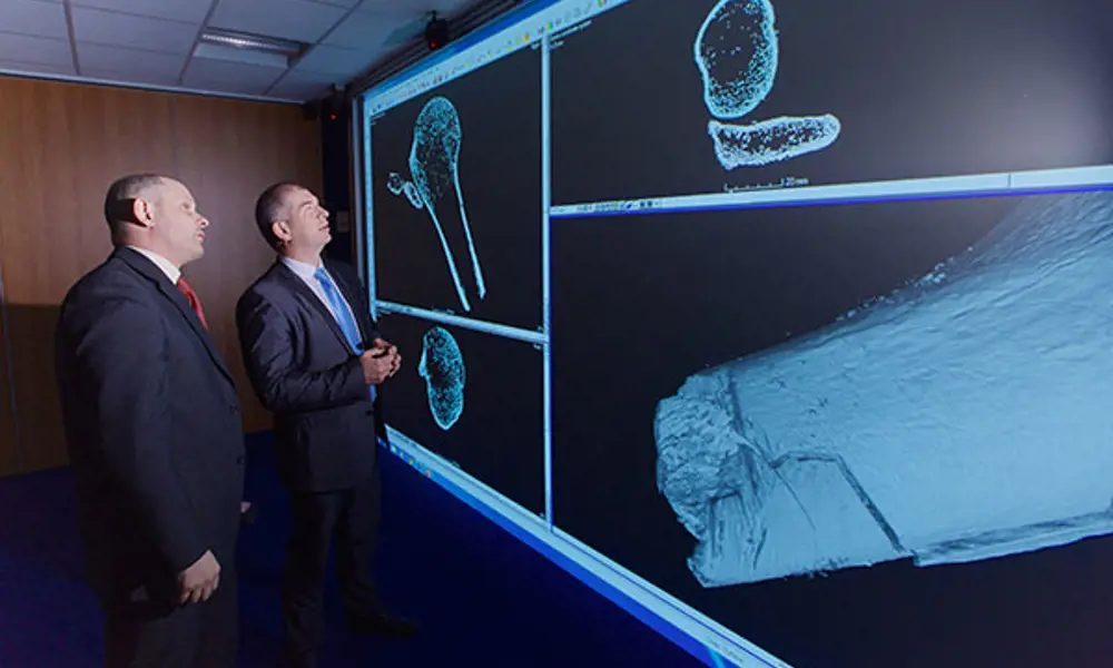
The range of distinctive marks resulting from bladed weapons and the unique characteristics of microscopic notches on bone. The top layer shows the physical bone section viewed under a microscope. The second layer is a 3D reconstruction from micro-CT data and the bottom level shows a corresponding 2D slice from a digital model © WMG
While 3D imaging and data processing can revolutionise the capture and presentation of virtual representation of crime scenes, this is but a part of the technological repertoire that is increasingly available in forensic analysis. Another tool that has the potential to make a significant impact is high-resolution X-ray computed tomography (CT), commonly referred to as micro-CT. University Hospital Coventry and Warwickshire (UHCW) hospital have carried out pioneering work on the use of medical CT for the digital documentation of post-mortem procedures.
However, micro-CT has the capability to characterise forensic specimens at up to a thousand times the resolution available in the healthcare scanning technology and can be used to identify and classify microscopic injuries – see CT setup.
Evaluating weapons and injuries
Micro-CT can also provide 3D analysis of objects involved in what are known as ‘sharp force injuries’. Home Office statistics suggest that nearly 40% of murders in the UK result from wounds caused by pointed objects or objects with sharp edges, including domestic knives, combat knives, scissors, axes and screwdrivers. Tool-mark analysis and weapon-wound matching, a developing area within forensic pathology, helps to establish the weapon class used to commit a homicide or to link a particular object to a specific crime.
Traditionally, analysis of sharp force injuries has been performed using established laboratory microscopy. This produces high-magnification 2D images that can be used to attempt to match the traumatic separation of tissue and skeletal damage with the unique characteristics of individual weapons. Although there have been successful cases, the ability to reliably match 2D digital images with real-world 3D objects has presented a significant challenge when presenting evidence in court. Micro-CT enables 3D microscopic evaluation of both weapons and injuries, particularly those on bone and hard tissue.
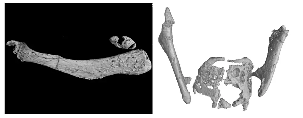
This research played an important part in a recent murder case. It helped to match items recovered near a body and to refine the search to look for a non-serrated weapon. A black lump resembling a large piece of coal was found in an oil drum in a suspect’s garden. Micro-CT scans revealed that it contained the top part of the victim’s humerus, fused inside a mass of molten debris. The bone had been sawn and snapped. After scanning the victim’s body parts, found in cases thrown into a canal, the recovered bone was found to be a perfect fit to another piece of bone belonging to the victim. The scan could show in minute detail – down to one 17,000th of a millimetre or half a hair’s breadth – the cuts on the bones.
a) Typical microscopic cracking to the rear quarter of the left hand greater horn of a hyoid bone from a case of strangulation with ligature. b) Fractures to calcified larynx cartilage consistent with manual strangulation © West Midlands Police
Micro-CT can also assist in the forensic analysis of strangulation. The human neck is a complex and delicate structure formed from numerous organs consisting of soft tissue, bone, calcified tissue, and cartilage. During a post-mortem, a victim may well exhibit visible external markers, including bruising or scratches, that potentially indicate the application of the forces required to cause death.
To help to establish the exact cause of death, further analysis of internal trauma would typically require the visual inspection of specimen dissections at the autopsy or further laboratory histology. These procedures are invasive and can destroy samples. Experience shows that injuries caused from strangulation are often subterranean in nature and hidden, or so small that they are extremely difficult to pick up with the human eye. Detection and imaging of these important indicators is usually beyond resolutions achievable through medical X-rays, conventional CT or magnetic resonance imaging.
Micro-CT can characterise commonly observed injuries, such as cracking to the hyoid bone or internal tearing to the larynx cartilages at a level of detail that allows detection and subsequent quantification. This process has proved successful for the West Midlands Police and was important in the presentation of evidence to the jury in five cases in the past two years. As a result, the force now uses micro-CT imaging of suspected strangulations as a part of its standard operating procedures.
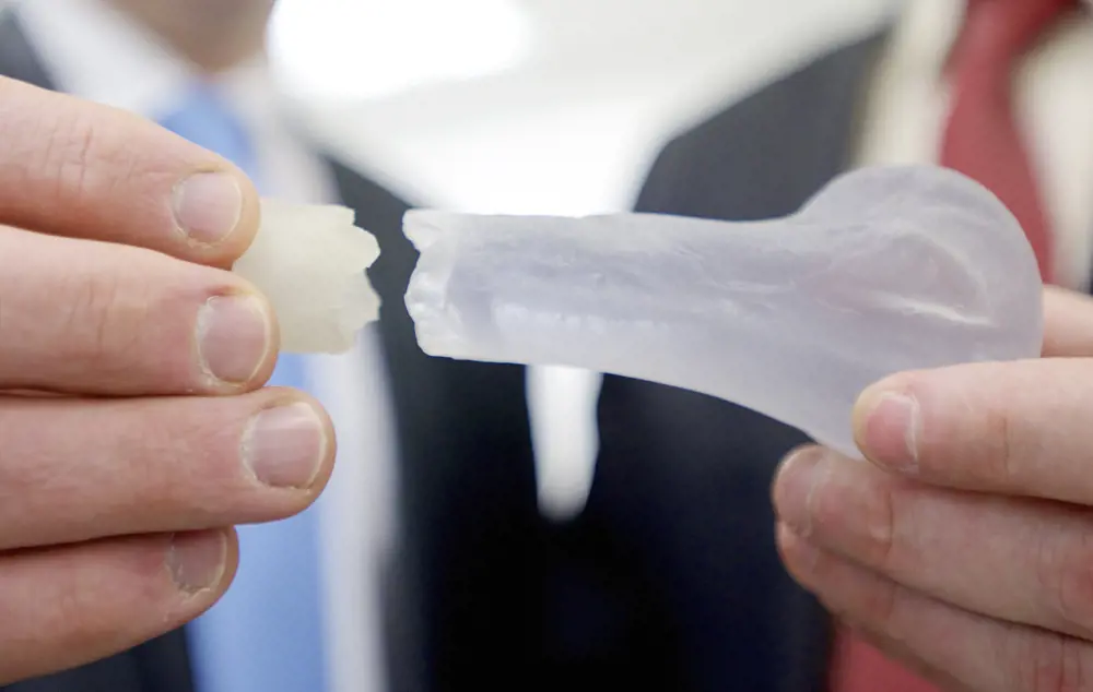
A 3D print of two parts of fractured bone from the same body. The section of bone on the right was recovered at the scene of the crime while the part on the left was found with the rest of the body at another location. WMG’s work helped prove that the two came from the same person © West Midlands Police
3D printing
Micro-CT and the ability to generate 3D images have also proved valuable in allowing forensic investigations to deploy another novel technology in the analysis of crimes and the presentation of evidence. 3D printing, also known as additive layer manufacturing, is now well-established for the manufacture of low volume components or parts with complex and unique geometry. The process of creating an object through the laying down of successive layers of material formed under computer control suits the generation of models for key pieces of evidence that can be presented at court.
One benefit of laser scanning and Micro-CT is that they generate data that are compatible with the stereolithography format that is accepted in 3D printers. This enables the generation of highly accurate physical representations of key pieces of evidence that can be produced at any scale.
Summing up
The examples described here show the increasing contribution that 3D imaging techniques and their application can bring to solving crimes and presenting evidence in court. The ability to create a photorealistic and accurate 3D digital archive of a location has a number of benefits to a forensic scene investigator, including the ability to analyse evidence long after the crime scene has been released from the criminal justice process. Another significant benefit is the ability to minimise confusion and to place a large quantity of detailed evidence in context to assist the jury in its determinations.
Cases that are particularly graphic and disturbing can prove particularly difficult when building a case for the prosecution. The potential to induce a decision based on emotive impact has resulted in key pieces of evidence being withheld from a jury. The ability to produce digital and physical prototypes that can be substituted for graphic photos allows the abstraction and subsequent sanitisation of potentially distressing evidence. The rigorous engineering processes that are used help to ensure the integrity of these models when challenged in court.
The ability to create a photorealistic and accurate 3D digital archive of a location has a number of benefits to a forensic scene investigator
These new engineering approaches to capturing and presenting 3D information are clearly influencing how the police solve crimes. The Home Office Pathologist, Dr Nicholas Hunt, has commented that the new techniques are complementary to current post mortem procedures and can provide additional help in establishing the cause of death. Evidence provided by the WMG team has supported a number of successful cases for the police.
It is a sign of the value that the police attach to these new techniques that in return for access to the technology and forensic experts, West Midlands Police is funding a three-year forensic PhD placement at WMG. This has increased the potential for the team at the University of Warwick to further adapt new technologies for forensic purposes and to solve serious crimes.
***
This article has been adapted from "Digital forensics", which originally appeared in the print edition of Ingenia 66 (March 2016).
Contributors
Professor Mark Williams PhD FIMechE leads the Product Evaluation Technologies and Metrology group at WMG, The University of Warwick. After gaining his PhD he went on to work in the automotive sector becoming a chartered engineer. He joined WMG in 2003 working on collaborative R&D projects and applied research funded through EPSRC, InnovateUK and the EU.
Keep up-to-date with Ingenia for free
SubscribeRelated content
Technology & robotics

When will cars drive themselves?
There are many claims made about the progress of autonomous vehicles and their imminent arrival on UK roads. What progress has been made and how have measures that have already been implemented increased automation?
Autonomous systems
The Royal Academy of Engineering hosted an event on Innovation in Autonomous Systems, focusing on the potential of autonomous systems to transform industry and business and the evolving relationship between people and technology.
Hydroacoustics
Useful for scientists, search and rescue operations and military forces, the size, range and orientation of an object underneath the surface of the sea can be determined by active and passive sonar devices. Find out how they are used to generate information about underwater objects.
Instilling robots with lifelong learning
In the basement of an ageing red-brick Oxford college, a team of engineers is changing the shape of robot autonomy. Professor Paul Newman FREng explained to Michael Kenward how he came to lead the Oxford Mobile Robotics Group and why the time is right for a revolution in autonomous technologies.
Other content from Ingenia
Quick read

- Environment & sustainability
- Opinion
A young engineer’s perspective on the good, the bad and the ugly of COP27

- Environment & sustainability
- Issue 95
How do we pay for net zero technologies?
Quick read

- Transport
- Mechanical
- How I got here
Electrifying trains and STEMAZING outreach

- Civil & structural
- Environment & sustainability
- Issue 95